
NCLEX Tip Subdural vs Epidural Hematomas Nclex, Nursing school
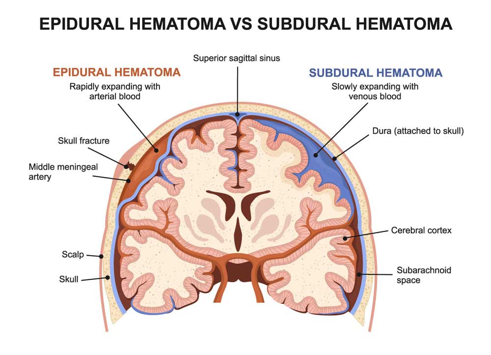
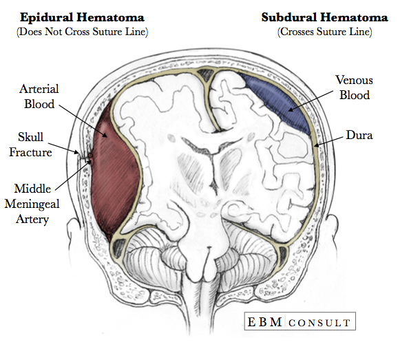
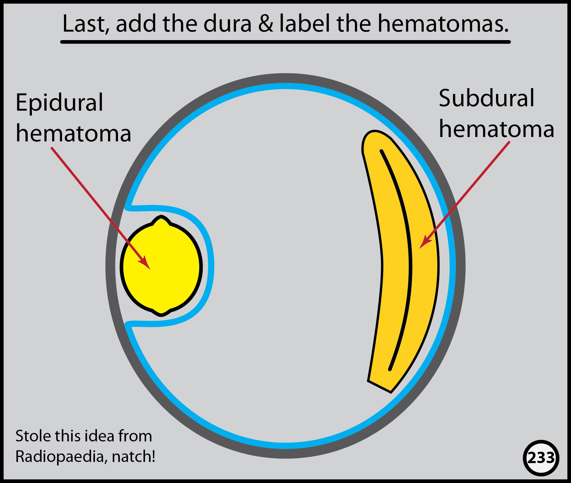
Epidural hematomas occur when an artery is injured and arterial blood accumulates between the dura and the calvarium. Do not cross suture lines because of the tight adherence of the dura to the calvarium and thus have a biconvex or elliptical appearance. The middle meningeal artery is classically involved, especially with a skull fracture.
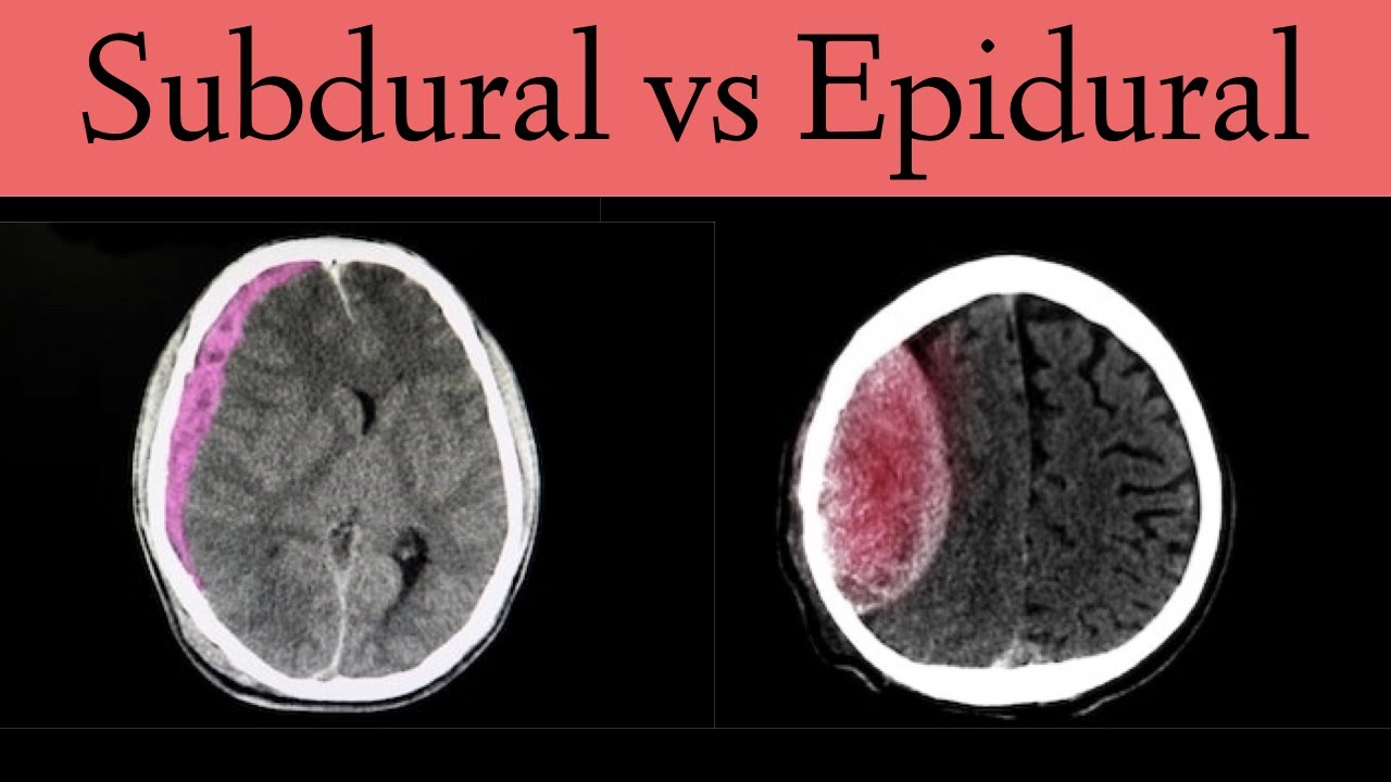
Subdural vs Epidural Hematoma/Hemorrhage [CT Scan Findings] YouTube
There are four types of ICH: epidural hematoma; subdural hematoma; subarachnoid hemorrhage; intracerebral hemorrhage; Epidural hematoma. A hematoma is a collection of blood, in a clot or ball.

Subdural vs epidural hematoma neurology nursing and medical notes Artofit
Subdural vs. epidural hematoma. Like a subdural hematoma, an epidural hematoma occurs when blood pools in the cranial tissue outside the brain. But in an epidural hematoma, the blood collects.

Head Trauma Trauma Orthobullets
Understanding the Difference Between Epidural & Subdural Hematomas neurosurgeons Although this is not always the case, head injuries like concussions In any case, the bleeding caused by an intracranial hematoma can form a mass that presses on the brain tissue and leads to a wide variety of potentially dangerous symptoms.

Subdural Hematoma VS Epidural Hematoma Intracranial Hemorrhages YouTube
TRAUMATIC EPIDURAL VS SUBDURAL HEMATOMA: Trauma to be brain can be associated with both epidural and subdural hematomas, among other injuries. Epidural bleeding occurs between the skull and dura; whereas subdural bleeding occurs between the dura and arachnoid.. An epidural hematoma is a neurosurgical emergency! Since bleeding is under.

Anatomy of EpiduralSubduralSubarachnoid space (from outside to inside
an epidural hematoma is a condition characterized by arterial bleeding developing in the potential space between the dura and the skull. head injury leads to a tear in the middle meningeal artery, leading to rapid filling in the epidural space, which compresses the parenchyma of the brain. the petrosal bone is thin, which can be easily.

SUBDURAL VS EPIDURAL HEMATOMA RADIOLOGY Wroc?awski Informator
Key Differences in Imaging. Location: The primary difference between the imaging of subdural and epidural hematomas is the location of the blood collection. Subdural hematomas are between the brain and the dura mater, while epidural hematomas are between the skull and the dura mater. Shape: Subdural hematomas typically appear crescent-shaped.
Subdural vs Epidural Hematomas Diller Law Personal Injury Law
An epidural is a procedure that involves injecting a medication — either an anesthetic or a steroid — into the space around your spinal nerves known as the epidural space. The goal of an epidural procedure is to provide pain relief (analgesia) or a complete lack of feeling ( anesthesia) for one region of your body, such as your legs or belly.

Epidural vs subdural Medical visual aids Pinterest
Symptoms include ongoing headache, confusion and drowsiness, nausea and vomiting, slurred speech and changes in vision. Subdural hematomas can be serious. See your healthcare provider if you have a head injury. Contents Overview Symptoms and Causes Diagnosis and Tests Management and Treatment Prevention Outlook / Prognosis Overview

Difference Between Subdural And Epidural
Pathology History and mechanism of injury Extradural hematoma The typical presentation is of a young patient involved in a head strike (either during sport or a result of a motor vehicle accident) who may or may not lose consciousness transiently.

Epidural Vs Subdural Hematoma / Epidural hematoma ALiEM Non
Epidural versus subdural hematoma From: Chapter 7, Traumatic Neuroemergency: Imaging Patients with Traumatic Brain Injury—An Introduction Copyright 2020, The Author (s)

Epidural vs Subdural Hematoma Medical graduate, Medical anatomy
Introduction Intracranial hemorrhage encompasses four broad types of hemorrhage: epidural hemorrhage, subdural hemorrhage, subarachnoid hemorrhage, and intraparenchymal hemorrhage. [1] [2] [3] Each type of hemorrhage is different concerning etiology, findings, prognosis, and outcome.
Anatomy Epidural vs Subdural Hematoma Image
Now, epidural hematoma is the collection of blood in the epidural space, which is the space between the dura mater and inner surface of the skull. In contrast, subdural hematoma is the collection of blood in the subdural space, meaning between the dura mater and the arachnoid mater. Let's start by looking at the physiology of the meninges.

Subdural vs Epidural Hematoma PDF Symptoms, Causes, CT, Radiology
Key Points. A spinal subdural or epidural hematoma is an accumulation of blood in the subdural or epidural space that can mechanically compress the spinal cord. Diagnosis is by MRI or, if not immediately available, by CT myelography. Treatment is with immediate surgical drainage. (See also Overview of Spinal Cord Disorders .)
Subdural Vs Epidural Hematoma RaeannArtin
So you can end up presenting with a headache and the nausea and the vomitting, and the cognitive changes for the subdural, a faster period of time. Those are sort of the 2 main differences between them, whereas a subarachnoid hemorrhage is caused by the arteries within the brain, and they run in the arachnoid space which was the middle layer.

Difference Between Epidural And Subdural Hematoma Pulptastic
Function. The epidural space serves as a cushioning and protective layer for the spinal cord and nerves. It is also the target for epidural injections used in pain management. On the other hand, the subdural space acts as a protective layer between the dura mater and the arachnoid mater, containing a small amount of fluid.